
SeeDB-Live/ACSF
This product allows fluorescent observation of neuronal activity while maintaining normal physiological functions by rendering the tissue transparent. Application of this product to acute brain slices has enabled, for the first time worldwide, non-invasive optical clearing of living tissue. This product is manufactured and sold by NACALAI TESQUE, INC. under a license from Kyushu University.
Features
- Transparent brain tissue with preserved normal neuronal activity
- Enabling deeper fluorescence imaging
- Non-cytotoxic
Application
Bright-field and Fluorescence imaging of living brain tissue
Acute brain slices (300 μm thick) were prepared from P5 mice. After perfusion with ACSF and SeeDB‑Live/ACSF, transmitted‑light images were acquired. In a separate experiment, olfactory bulb slices from P13 Thy1‑GCaMP6f mice were imaged using confocal microscopy.
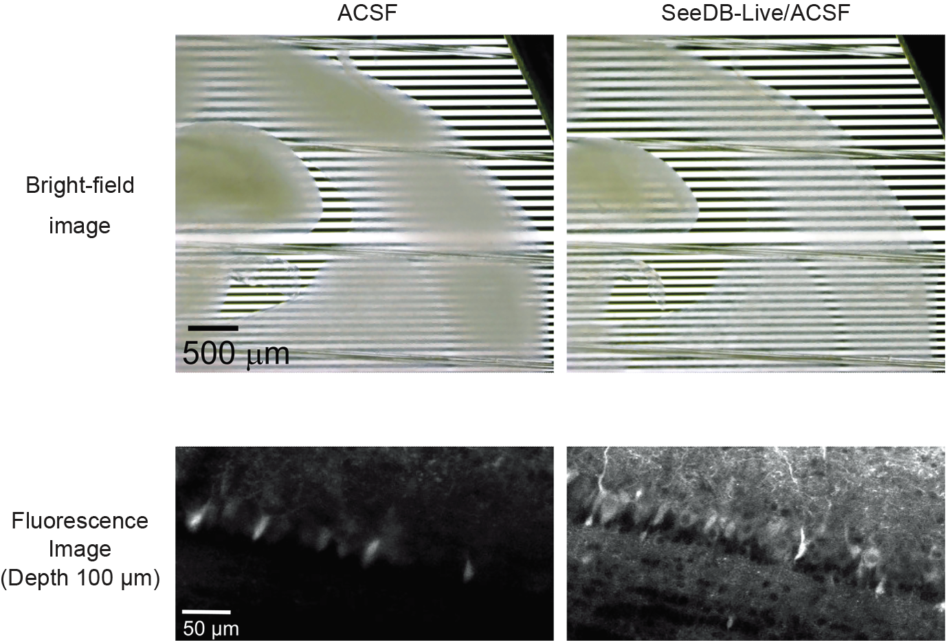
These data were provided by Professor Takeshi Imai and Assistant Professor Shigenori Inagaki, Faculty of Medical Sciences, University of Kyushu.
Calcium imaging in acute olfactory bulb slice from Thy1-GCaMP6f mice
Acute olfactory bulb slices from Thy1-GCaMP6f mice were optically cleared using either SeeDB-Live/ACSF or ACSF and imaged by two-photon microscopy. The results demonstrated that SeeDB-Live/ACSF enables deep imaging of live tissue while preserving normal cellular function.
| ACSF | SeeDB-Live/ACSF | ||
| Depth 90 μm | |||
| Depth 150 μm | |||
| Depth 200 μm |
These data were provided by Professor Takeshi Imai and Assistant Professor Shigenori Inagaki, Faculty of Medical Sciences, University of Kyushu.
References
Isotonic and minimally invasive optical clearing media for live cell imaging ex vivo and in vivo
Inagaki S, Nakagawa-Tamagawa N, Huynh NZ, et al. Nat Methods. 2026.
DOI : https://doi.org/10.1038/s41592-026-03023-y
Downloads
SeeDB-Live/ACSF  (PDF 1.8 MB)
Ordering Information
| Product name | Grade | Storage | Catalog number | PKG size | Price |
|---|---|---|---|---|---|
| SeeDB-Live/ACSF | SP | Refrigerate | 23041-44 | 100 mL |
